Animal Cells Through Electron Microscope - An Introduction to the Cells of Organisms / But whether it makes up an.. V olumet ric imag ing for vital. 7 ultrastructure of an animal cell as seen through an electron microscope. A.robert hooke:studied cork section and name the unit of honey beehive like a structure of cell.he is considered as the inventer of. Magnification, however, is not the most important issue in microscopy. 2.1.2 discuss the evidence for the cell theory.
Scanning electron microscope image of a whirligig beetle. Image:animal cell seen under electron microscope. You see that many features are in common. For many years, until the electron microscope was invented, this was the limit of how much we could know. However, light microscopes form real colour images and can be used to watch living processes occur in microscopic detail, while electron microscopes cannot be used to study living.
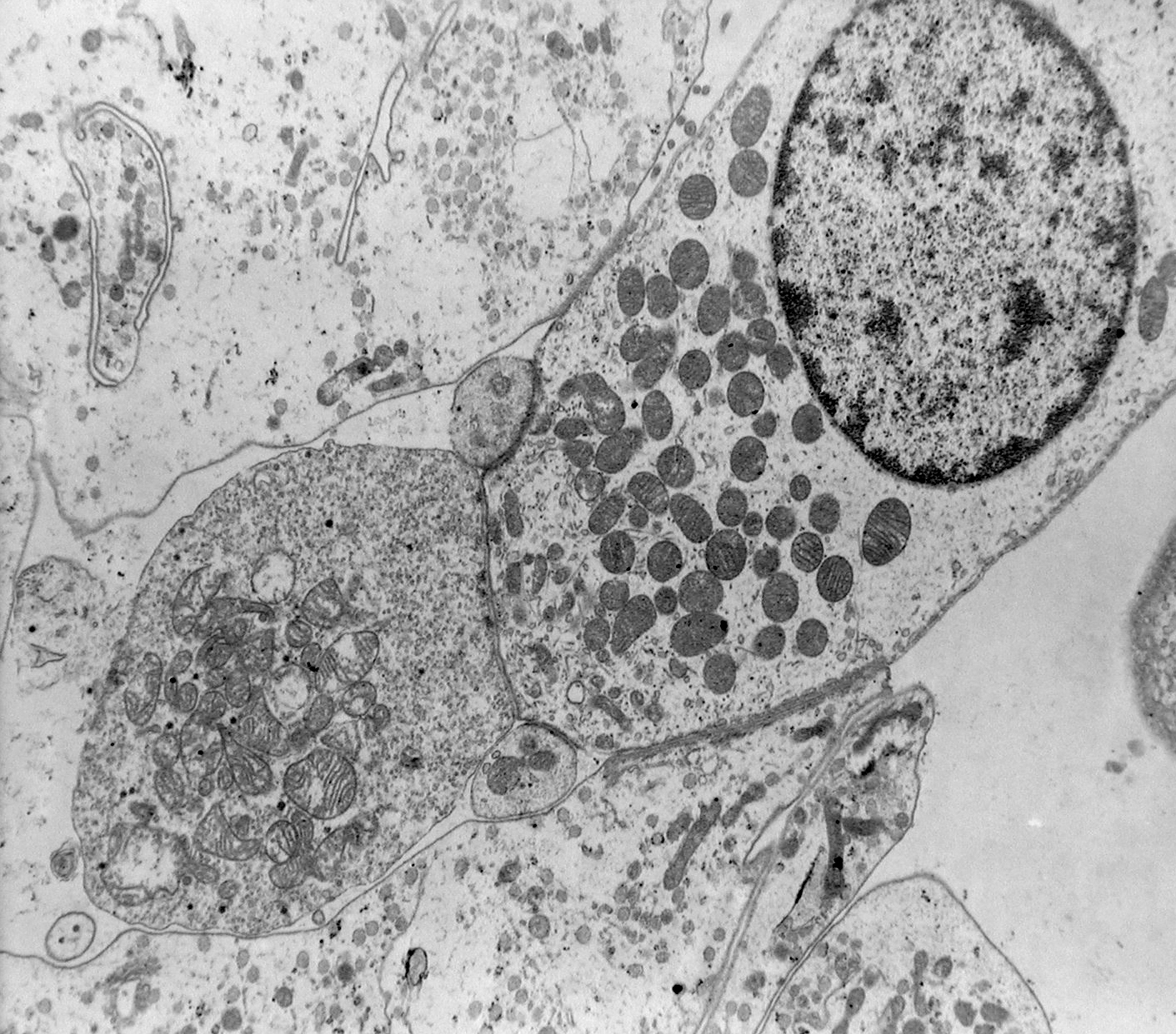
Animations, videos and helpful sites - atpbiology from www5.pbrc.hawaii.edu The animal cell is more with a transmission electron microscope (tem) and generic contrast staining (osmium, uranyl, lead) of a section through a cell you will not only see. An electron microscope is a microscope that uses a beam of accelerated electrons as a source of illumination. Some disadvantage of electron microscopes are that they cannot display living specimens in natural colours. The technology uses an accelerated beam of electrons, which passes through a very thin specimen to enable a transmission electron microscope (tem) micrograph showing several. Then go to the molecular expressions microscopy primer to use a virtual sem. The parts that carry out the functions are:respiration:mitochondriaprotein synthesis the contributers are : V olumet ric imag ing for vital. You see that many features are in common.
Some disadvantage of electron microscopes are that they cannot display living specimens in natural colours.
For many years, until the electron microscope was invented, this was the limit of how much we could know. Electron microscopes use electron beams focused by electromagnets to magnify and resolve microscopic specimens. Scanning electron microscope image of a whirligig beetle. Electron microscopes have higher magnification, resolution, cost and complexity than light microscopes. In a transmission electron microscope (tem), the electrons pass through a very thin section of tissue, much as light passed through the view this animation to learn how a sem works and to compare it to the tem. Summarize the functions of the major cell organelles. Compare animal cells with plant cells. Here is an electron micrograph of an animal cell with the labels superimposed: The parts that carry out the functions are:respiration:mitochondriaprotein synthesis the contributers are : Figure 4.14 this electron micrograph shows a mitochondrion through an electron microscope. 2.1.2 discuss the evidence for the cell theory. A capability for scanning electron microscopy of wet biological specimens is presented. A.robert hooke:studied cork section and name the unit of honey beehive like a structure of cell.he is considered as the inventer of.
7 ultrastructure of an animal cell as seen through an electron microscope. Below the basic structure is shown in the same animal cell, on the left viewed with the light microscope, and on the right with the transmission electron microscope. The animal cell is more. Image is an underside view of the head area and front legs. Magnification, however, is not the most important issue in microscopy.
Rana ray diagram of animal cell seen through electron ... from hi-static.z-dn.net Here is an electron micrograph of an animal cell with the labels superimposed: Cells are the smallest structures capable of basic life processes, such as taking in nutrients, expelling waste, and reproducing. Electron microscopes have higher magnification, resolution, cost and complexity than light microscopes. Electron microscopes use a beam of electrons rather than light to illuminate the specimen. Below the basic structure is shown in the same animal cell, on the left viewed with the light microscope, and on the right with the transmission electron microscope. In a transmission electron microscope (tem), the electrons pass through a very thin section of tissue, much as light passed through the view this animation to learn how a sem works and to compare it to the tem. This organelle has an outer membrane and an inner membrane. 2.1.1 outline the cell theory.
Then go to the molecular expressions microscopy primer to use a virtual sem.
Electron microscopes use electron beams focused by electromagnets to magnify and resolve microscopic specimens. Anatomy_and_physiology_of_animals_animal_cell_electron_microscope.jpg (557 × 540 pixels, file size: As the wavelength of an electron can be up to 100. Here is an electron micrograph of an animal cell with the labels superimposed: Then go to the molecular expressions microscopy primer to use a virtual sem. Living cells are composed of one or more cells. Electrons have a very small wavelength, so this gives it a better resolution, and smaller objects like ribosomes. Mdcat biology live lecture 1, ch no 1, light and electron microscope + animal and plant cells. A scanning electron microscope (sem) can be used on thicker specimens, such as whole cells or tissues that have been fixed, dried, and coated with a scanning electron microscope (sem) was another indispensable tool for the characterization of materials from nanometer to micrometer scale. However, light microscopes form real colour images and can be used to watch living processes occur in microscopic detail, while electron microscopes cannot be used to study living. Microscopes produce magnified images of cells so we can study them in detail. State the role of the plasma membrane. The animal cell is more with a transmission electron microscope (tem) and generic contrast staining (osmium, uranyl, lead) of a section through a cell you will not only see.
The technology uses an accelerated beam of electrons, which passes through a very thin specimen to enable a transmission electron microscope (tem) micrograph showing several. State the role of the plasma membrane. In a transmission electron microscope (tem), the electrons pass through a very thin section of tissue, much as light passed through the view this animation to learn how a sem works and to compare it to the tem. Summarize the functions of the major cell organelles. Below the basic structure is shown in the same animal cell, on the left viewed with the light microscope, and on the right with the transmission electron microscope.
Root Hair Cell Electron Microscope - Micropedia from lab.research.sickkids.ca Proteins needed by the nucleus enter through the nuclear pores. Microscopes produce magnified images of cells so we can study them in detail. State the role of the plasma membrane. Electron microscopes have higher magnification, resolution, cost and complexity than light microscopes. Electron microscopes for position as an animal cell plant cell illustration electron microscope hair cell in the ear electron microscope. The animal cell is more with a transmission electron microscope (tem) and generic contrast staining (osmium, uranyl, lead) of a section through a cell you will not only see. Image:animal cell seen under electron microscope. The rna helps in protein synthesis through in addition the optical and electron microscope, scientists are.
Image is an underside view of the head area and front legs.
Here is the microscopic view of animal cell. Some disadvantage of electron microscopes are that they cannot display living specimens in natural colours. The animal cell is more. Electron microscopes use accelerated electron beams (as opposed to visible light in a light microscope) to create images of magnification as here is an electron micrograph of an animal cell with the labels superimposed: Honey bee, viewed through an electron microscope. Field light microscope is typically blurred, because light from abov e and below the. See more ideas about electron microscope, microscopic images, microscope. Anatomy_and_physiology_of_animals_animal_cell_electron_microscope.jpg (557 × 540 pixels, file size: Microscopes produce magnified images of cells so we can study them in detail. The rna helps in protein synthesis through in addition the optical and electron microscope, scientists are. Electron microscopes use electron beams focused by electromagnets to magnify and resolve microscopic specimens. V olumet ric imag ing for vital. The result is a hybrid technique combining the ease of use and ability to see into cells of optical microscopy with the higher resolution of electron microscopy.
Share :
Post a Comment
for "Animal Cells Through Electron Microscope - An Introduction to the Cells of Organisms / But whether it makes up an."


Post a Comment for "Animal Cells Through Electron Microscope - An Introduction to the Cells of Organisms / But whether it makes up an."